Diagnosis of endometriosis
The diagnosis begins with a detailed discussion with your doctor and a summary of all the findings.
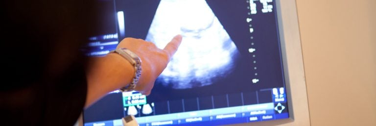
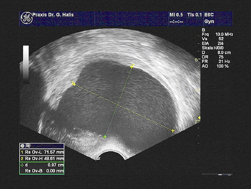
It would be good if you could bring all documents such as surgery reports, doctor’s letters, findings, etc. with you to the consultation. We will send you a patient questionnaire in advance, which you can fill out at home and email to us. After the consultation, a careful palpation and ultrasound examination will take place.
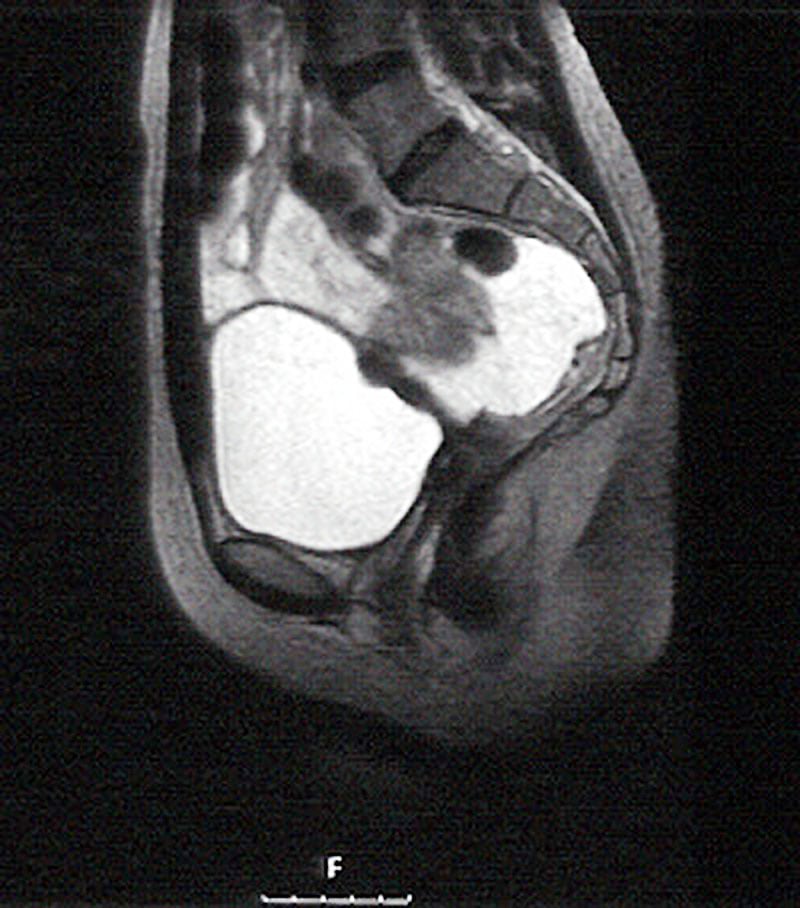
Depending on the symptoms and the question, an examination with other procedures may also be useful, e.g. a colonoscopy or imaging procedures such as magnetic resonance imaging.
Depending on the symptoms and the indication, assessment by means of other procedures may be advisable. These may include colonoscopy or diagnostic imaging procedures such as magnetic resonance imaging (MRI).
The final proof of endometriosis, however, can only be provided by a laparoscopy. During this surgical procedure under anaesthesia, the stage of the disease is determined and the endometriosis is surgically treated. If there is a desire to have children, it should be checked simultaneously whether the fallopian tubes are open or whether there are abnormalities in the uterus.