Diagnosis of fibroids
Non-symptomatic fibroids are often discovered as an incidental finding, e.g. during a gynaecological check-up. If symptoms are present, the doctor can specifically look for possible fibroids.
How do doctors recognise fibroids?
Overview of the most important diagnostic steps for fibroids:
Anamnesis (medical history)
In the anamnesis, the doctor takes the personal medical history. In addition, he asks about current and past physical complaints. A regularly kept cycle calendar helps the doctor to get an overview of the occurrence of menstrual symptoms, length of bleeding and intermenstrual bleeding. In addition, the doctor will ask about previous treatments and the use of medication (e.g. the pill). The family history is also important, for example, whether the mother or sisters have already suffered from menstrual problems or fibroids.
Palpatory examination
During bimanual palpation, the gynaecologist palpates the pelvis and assesses the size, position, mobility, consistency and dolence (painfulness) of the uterus and adnexa.
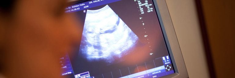
Ultrasound examination
Ultrasound examination through the vagina (vaginal sonography) or via the abdomen (abdominal sonography) is a very effective method of examining the female reproductive organs. If fibroids are present, the gynaecologist can determine the number and location of the fibroids more precisely and measure their size. This technique also makes it possible to observe fibroids over a longer period of time and to assess the blood flow patterns with the help of Doppler sonography.
In particularly severe cases, other methods can help to diagnose fibroids:
Uteroscopy
Hysteroscopy was first developed as a method to examine the inside of the uterus. Later it became surgical hysteroscopy, which can be used to remove submucous fibroids. Much depends on the size and location of the fibroids and the experience of the surgeon.
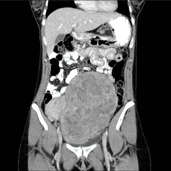
CT/MRI
Myomas are sometimes discovered as an incidental finding when a CT or an MRI has been performed for other reasons. Because of the better tissue specificity and the lack of radiation exposure, an MRI should only be performed if necessary.